Advanced Diagnostics
Precise, technology-driven exams to identify what’s causing your dry eyes.
Defining the Underlying Cause
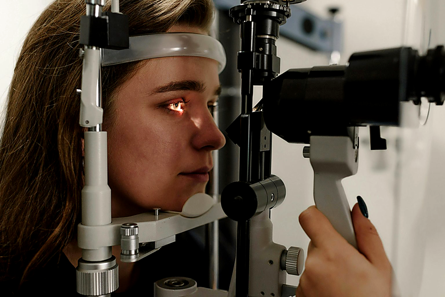
We use advanced imaging and analysis to look beyond your symptoms and identify the specific dysfunctions causing your dry eyes. The results of these structured diagnostic exams guide your treatment plan – that’s what makes it work.
Diagnostic Tests
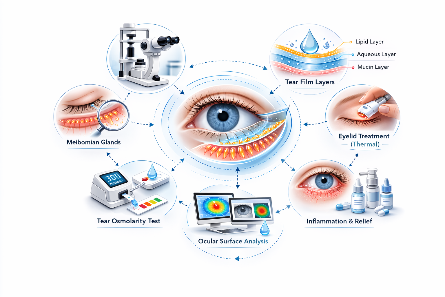
Given the complexity of dry eye disease, a single test isn’t enough — multiple measurements are required.
NIBUT (Non-Invasive Breakup Time)
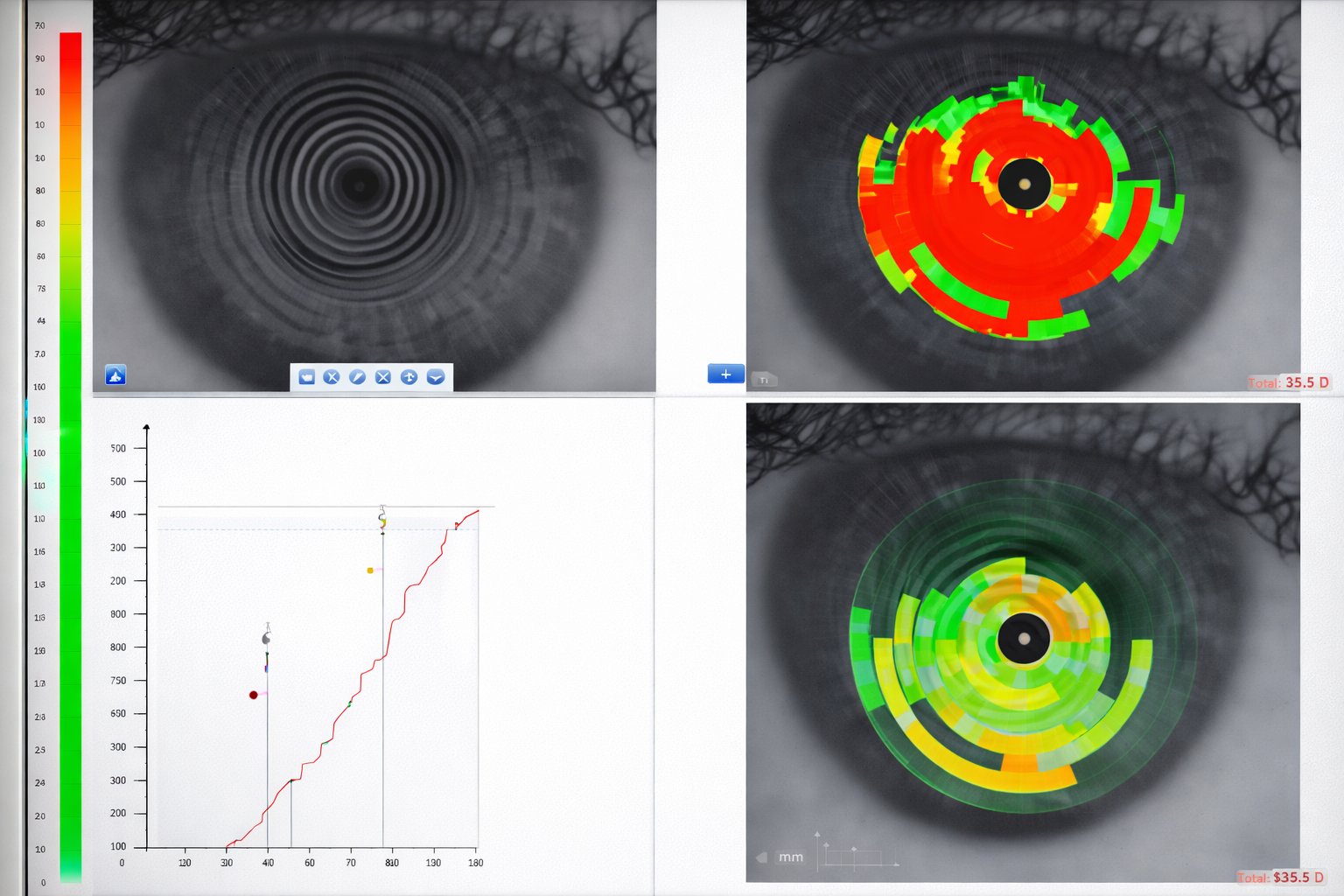
Measures how long your tear film remains stable after a blink — without dyes or direct contact.
- Dye-free
- Completed in seconds
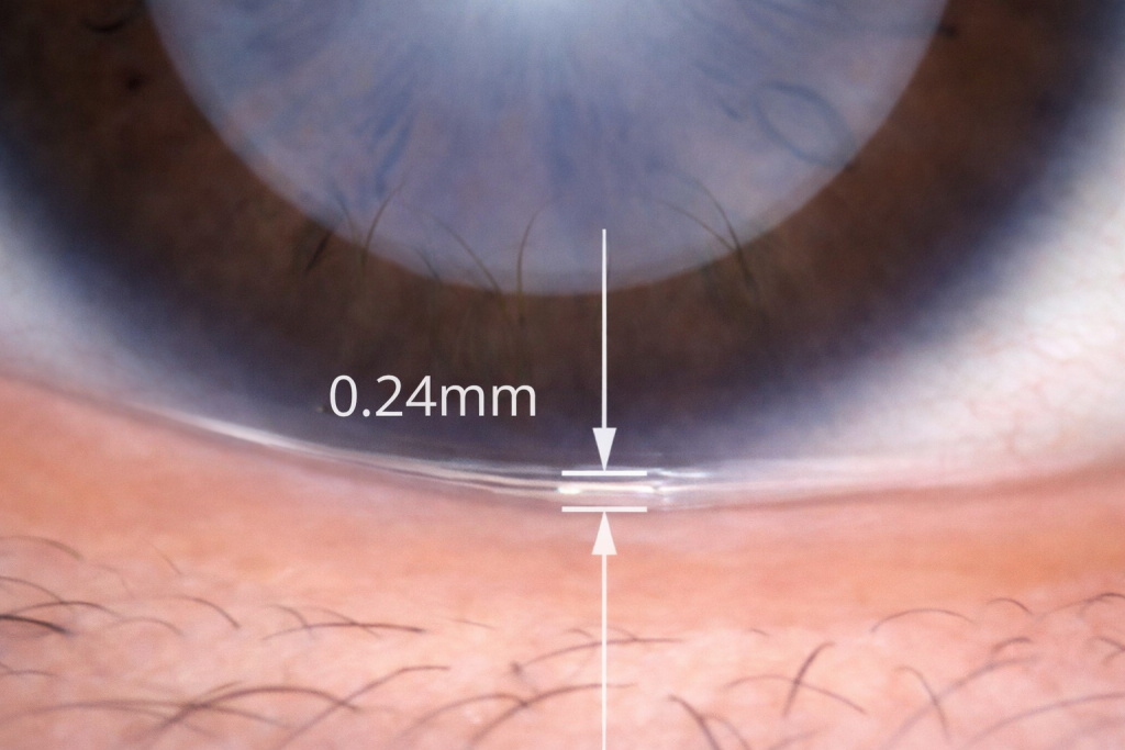
Tear Meniscus Height (TMH)
Measures the volume of tears along your eyelid margin to assess whether you’re producing enough.
- Contact-free
- Rapid measurement
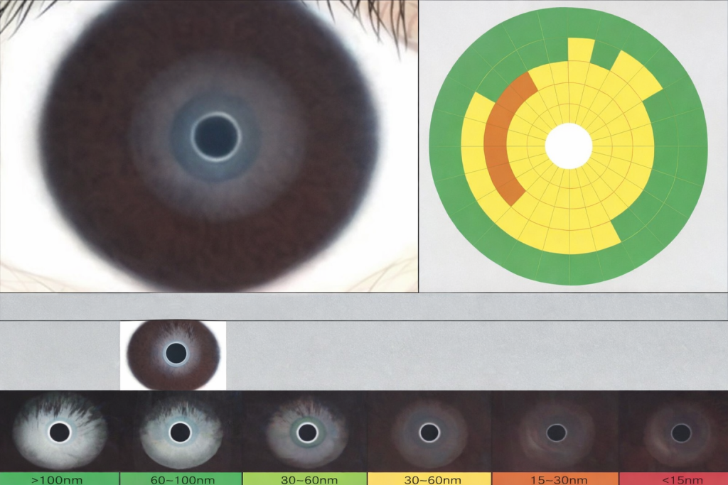
Lipid Layer Analysis
Evaluates the oily outer layer of your tear film, which prevents evaporation. Thinning here often points to meibomian gland dysfunction.
- 1–2 minutes
- Performed during natural blinking
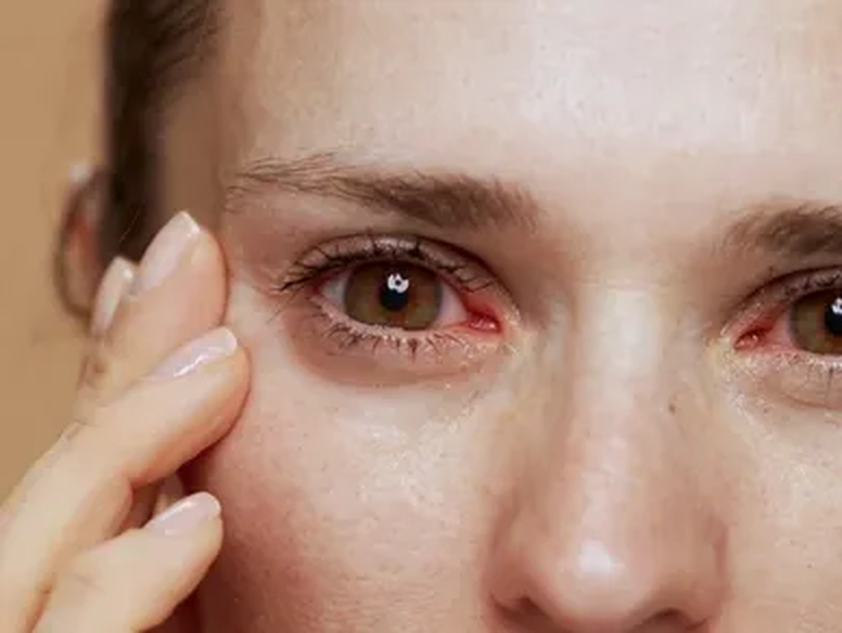
Eye Redness Analysis
Maps inflammation across the conjunctiva and sclera to assess severity and track changes over time.
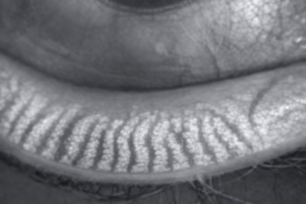
Meibomian Gland Imaging
Visualizes the structure and health of the glands responsible for producing the lipid layer. Gland loss is often irreversible — early detection matters.
Gland Openings
The meibomian glands release oils through tiny openings along the eyelid margin with every blink. When these become blocked, the oils can’t reach the tear film, leading to evaporative dry eye. If blockages persist, gland loss can become permanent. Assessing these openings is a key step in diagnosis and guides the initial therapy.
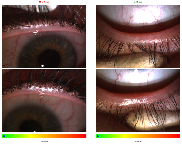
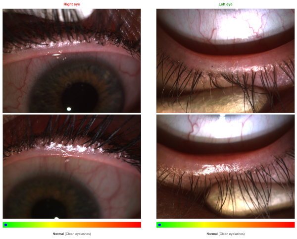
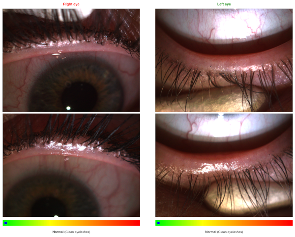
Eyelashes and Lid Margin Assessment
Eyelid inflammation (blepharitis) often involves a combination of bacterial overgrowth, meibomian gland dysfunction, and in some cases rosacea or Demodex mite infestation. High-resolution imaging captures crusting, swelling, redness, and possible deposits of demodex mites (collarettes) — all of which play a key role in your treatment plan.
Lid Margin Vessel Analysis
Dilated blood vessels along the lid margin (telangiectasias) may indicate rosacea-related dry eye disease and help refine your diagnosis.
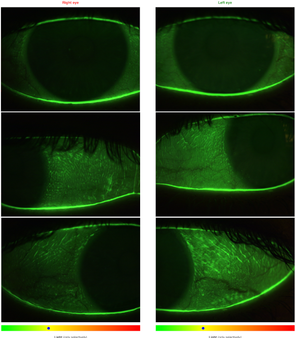
Conjunctival and Corneal Staining
A harmless fluorescein dye reveals dry patches and surface damage on the cornea and conjunctiva. The pattern and severity of staining indicate how advanced the condition is, help identify the cause, and provide a clear baseline to measure treatment progress.
All diagnostic procedures within the Smart Dry Eyes pathway are non-invasive and painless
Start Your Evaluation
Take the first step — begin a guided screening and get matched with the right care provider.